+40.232.261.950
office@dacorumgrup.ro
Bd. Chimiei, no. 89, Iași, România

Furnizori > Philips > Patologie digitală > Sistem de management al imaginilor (IMS)

Image Management System (IMS)

PHILIPS

Digital Pathology Visualization Software

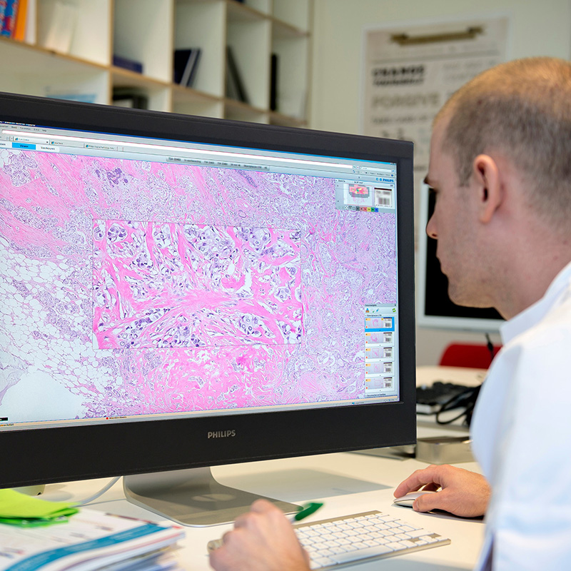
  • Integrated digital platform for the management and processing of digitized images for anatomopathology and histopathology
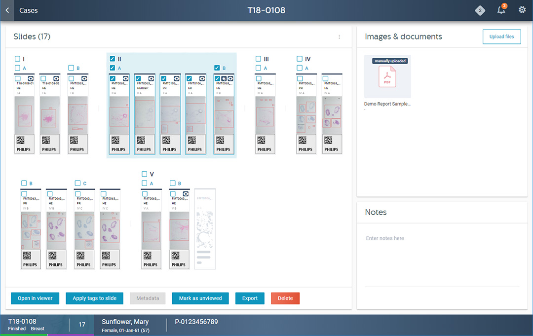
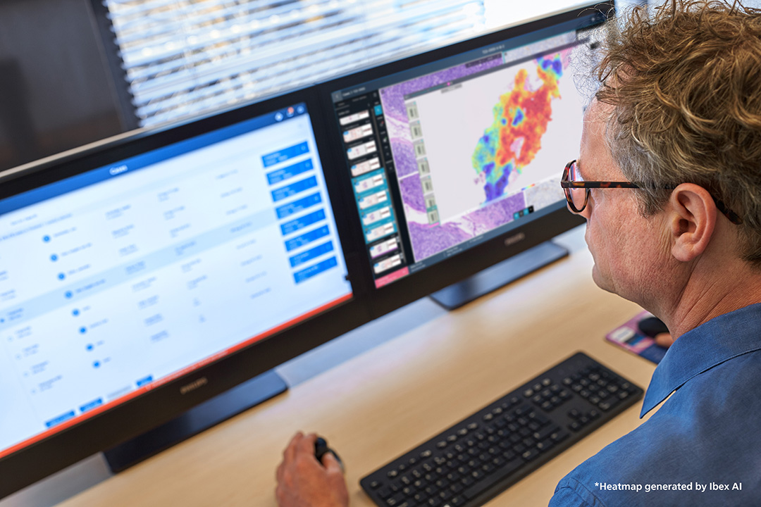
  • Modern interface with clear case organization and intuitive slide visualization
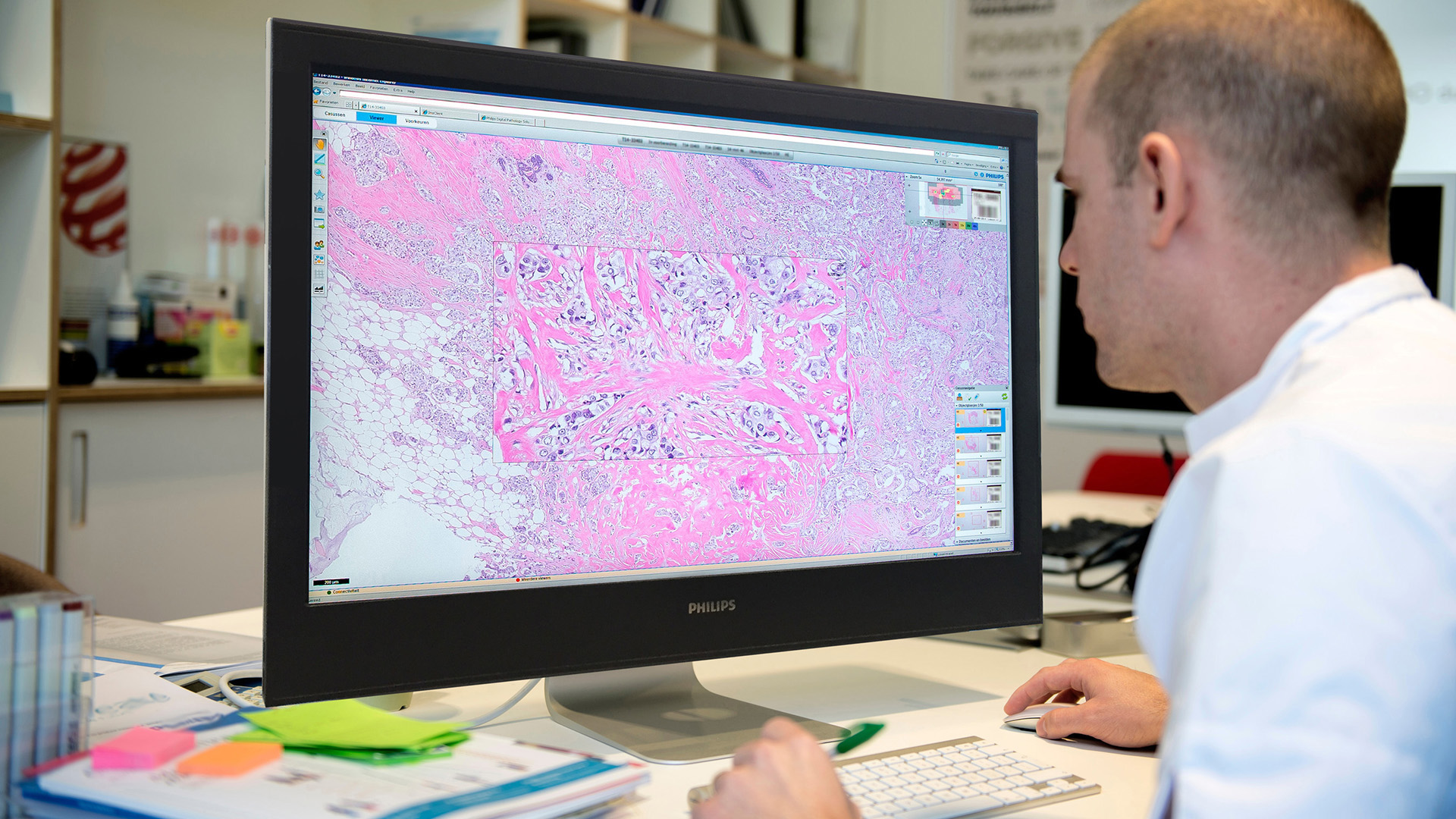
  • Digital slide tray with instant preview and visual indicators of completeness
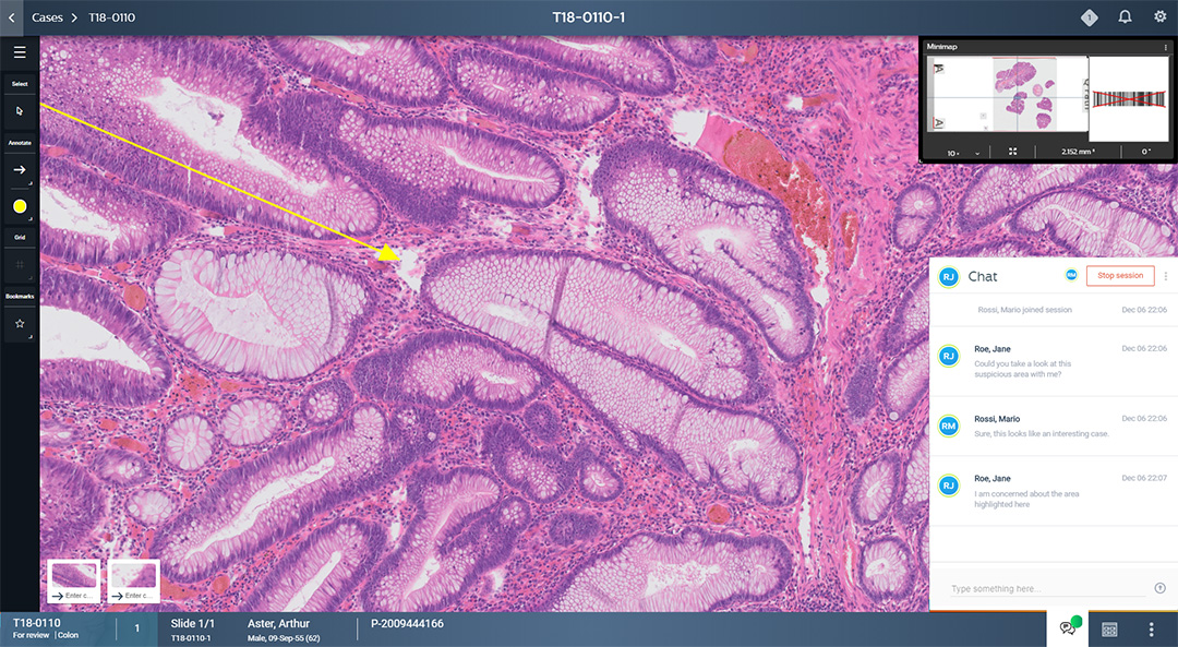
  • Advanced viewer with measurement and annotation tools for tissue analysis
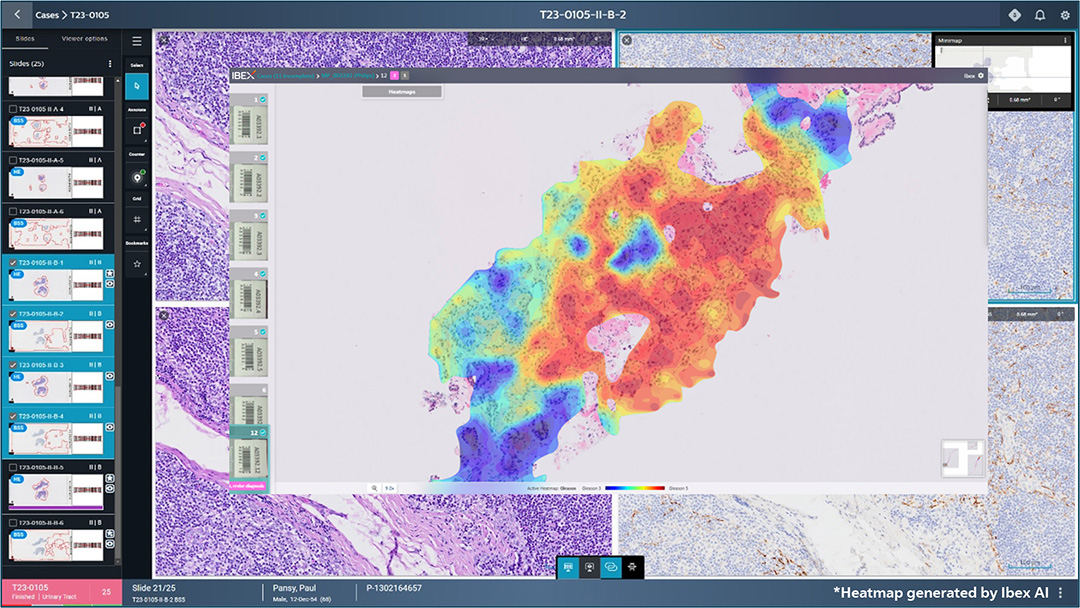
  • Native AI integration, including analysis status and direct access to Ibex AI
  • Objective and reproducible AI results that increase diagnostic accuracy and efficiency
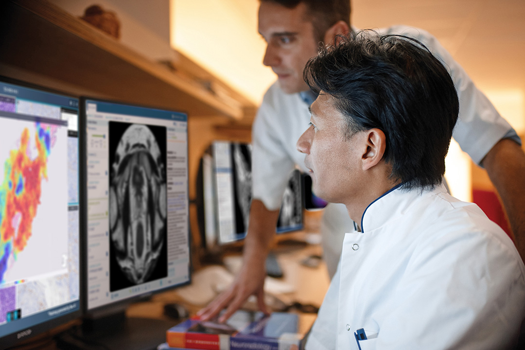
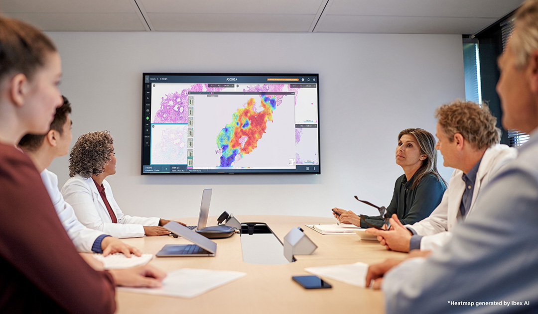
  • Structured and scalable digital workflow with support for real-time collaboration
  • Remote access to cases and images with optimized performance for distributed work
  • Intelligent worklist management with sorting, prioritization, and quick view
  • Full interoperability with multiple scanners and vendor-neutral compatibility
  • LIS integration with automatic barcode-based case organization
  • Clinical standards support, including DICOM JPEG2000 export
  • Scalability for labs of any size, including multi-site networks

Efficient workflow management

Organized personal work list that visually indicates the workload.

All the information about the case in one place

The digital blade tray with case details provides a clear overview of the case.

Advanced Tissue AnalysisFully equipped viewer that offers multiple tools for working with tissues.

Patient data is always accessible via barcode, without the need to check the labels on the bottle. After scanning, cases are automatically organized into patient-level folders (based on LIS interoperability).

Modern interface, optimized for clarity and speed

The IMS interface is designed to provide a complete picture of the workload and active cases. The list of cases is clearly structured, with visible status, quick filters and intuitive navigation. The digital slide tray replicates the physical organization of the slide tray, facilitating the structured analysis of blocks and sections.

Instant previews help check tissue quality before opening in the viewer. Icons for annotations, viewed areas, completeness, or the need for additional sections reduce errors and speed up case selection. The design is efficiency-oriented, so specialists spend less time navigating and more time analyzing.

Built-in AI analytics for augmented diagnostics

IMS acts as a central point for the integration of artificial intelligence applications, especially Ibex AI. The system instantly shows the results of the AI analysis and the progress of each case, allowing one-click launch to the AI platform without interruptions in the stream.

Ibex AI automatically highlights relevant areas, quantifies biomarkers and provides reproducible diagnostic scores, supporting a safer clinical decision. Proven efficiency in use includes

This integration positions IMS as a system that not only stores and displays images, but raises the diagnostic performance level of the entire laboratory.

Efficient and scalable digital workflow

IMS supports a completely digital journey, from the reception of the slides to the final analysis. The worklists are logically structured and visually reflect the status of each slide, including quick views, slide placeholders, and sorting by priority levels.

The preview of slides helps to verify the completeness of the tissues, and the real-time collaboration allows immediate consultation between specialists. The system can also be accessed remotely, allowing pathologists to review, annotate, and collaborate on cases regardless of location without loss of performance.

The structure supports scaling for multiple sites, connecting the flows of the entire network into a coherent digital ecosystem.

Interoperability and vendor-neutral compatibility

IMS is built as an open, flexible, and vendor-agnostic platform that can be integrated with scanners from Leica, Hamamatsu, 3DHISTECH, Glissando, and others. Laboratories can maintain their existing infrastructure, without technological bottlenecks or forcing migration to a single manufacturer.

Full integration with LIS ensures automatic organization of cases based on barcode, and lossless DICOM JPEG2000 export support allows data exchange according to clinical standards.

This open approach makes IMS suitable for both small laboratories and extensive networks or institutions that want to adopt AI and digital pathology without supply-related barriers.

  • 27% increased efficiency
  • 37% increased productivity
  • Reduction of analysis time by 43 hours in prostate cases
  • Used in 38 laboratories around the world