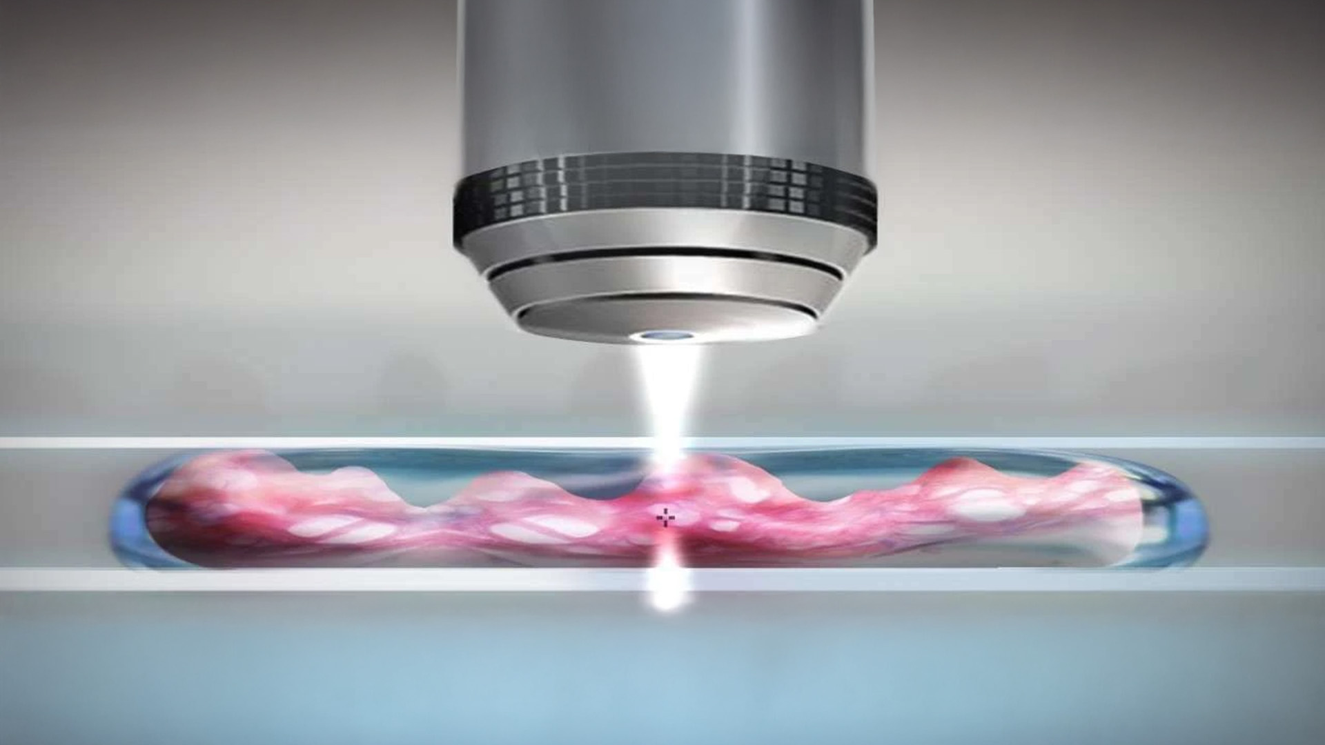
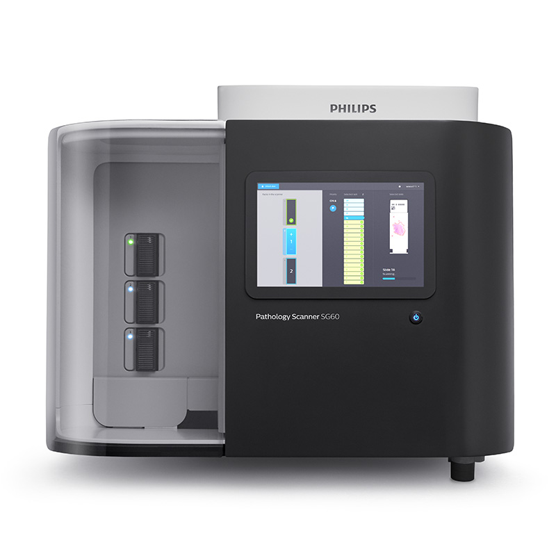
Furnizori > Philips > Patologie digitală > Pathology Scanner SG60


Pathology Scanner SG60
PHILIPS
-
Brightfield scanner
-
Designed to meet the needs of laboratories with a medium case volume
-
Tissue shape detection – the scanner adapts the scanning region to the tissue shape, resulting in shorter scan times and reduced storage requirements
-
Per-slide calibration check – for each scan, a calibration check is performed. Only if the check fails will the scanner automatically initiate recalibration. This ensures that every slide is scanned with a calibrated system and that recalibration occurs only when truly necessary
-
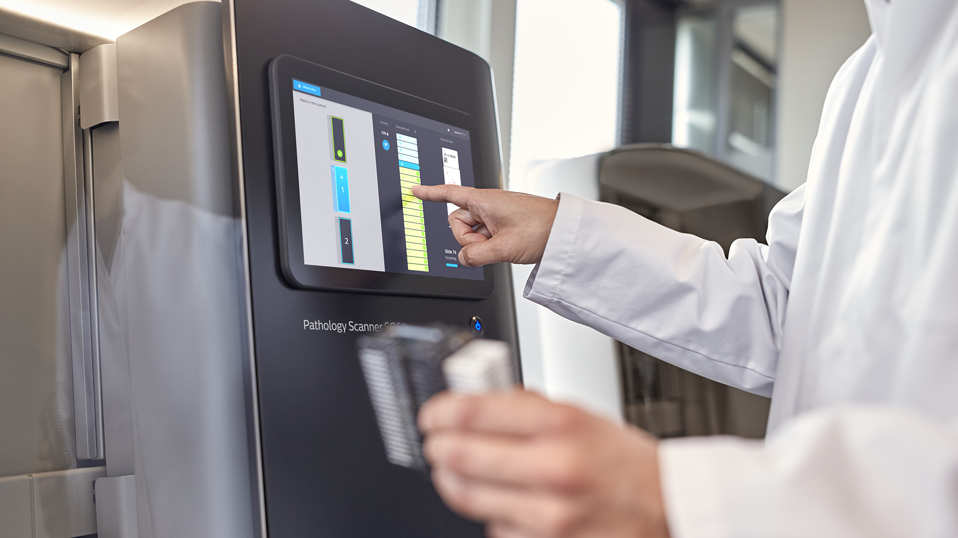
Easy to use – the user only needs to load and unload the slides; all other steps are fully automated
-
Priority flag assignment – the scanner allows a priority flag to be assigned to a rack. When a priority flag is assigned, the rack is moved to the front of the scanning queue, and the resulting slide images from the priority rack are sent to the IMS with a priority flag
-
Scan process status monitoring – can be checked via a color-coded LED (one LED for each rack), through the user interface, and via remote access
-
Can accommodate 3 racks – when fully loaded, it can store up to 60 slides
-
Multi-layer scanning – hardware prepared for 3D technology
High quality images
The scanner automatically produces clear digital images at 40x magnification (0.25 μm/pixel), essential for digitizing the diagnostic workflow and using advanced image analysis algorithms.
The scanner delivers high-quality images using a standardized and fully automated scanning process. Each slide is scanned at 40x equivalent magnification (0.25 um/pixel). This results in clear, high resolution images, essential for digitizing the primary diagnostic workflow and a key factor in the use of image analysis algorithms.


Uninterrupted Process
The ultra-fast scanner operates simply with a “load and go” system, automatically digitizing slides. Intelligent algorithms detect tissue, barcodes, protective glass and maintain continuous autofocus throughout the process.
Using the ultra-fast scanner requires simple ‘load and go’ operation. Anyone in the lab can load slides into the scanner and the digitizing process will continue automatically. This automated process is supported by numerous algorithms for tissue, barcode and coverglass detection, as well as continuous auto-focus algorithms used throughout the scan.
In just 43 seconds, a slide with a reference scanning area of 15×15 mm² is scanned with a resolution equivalent to a 40x magnifying objective with a numerical aperture of 0.75 (including handling and pre-scanning, the total scanning speed is ≤ 62 seconds).